
Osteochondroma of Rib (benign)
IMAGE 1. Specimen of a typical or common rib osteochondroma, resected. Its size is usual or average.
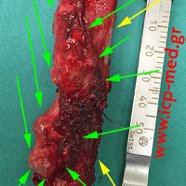
IMAGES 2–13:
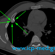
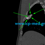
A Sizeable osteochondroma, measuring 6.7 × 4 × 2.2 cm. Its shape is irregular as it were to consist of two components. The tumour is located in the 4th rib (right–sided, antero–lateral aspect of it) of a 26–yo male athlete, a non–smoker. Radiological incidental finding.
Preoperative imaging investigations (CXR, CT, MRI scans, 99mTc bone scan with a SPECT / CT study) and photographs of the resected specimen.
Green arrows: the Tumour (osteochondroma).
Yellow arrows: the normal or healthy–appearing part of the rib (as well as the surgical resection margins)
IMAGES 2–13:
A Sizeable osteochondroma, measuring 6.7 × 4 × 2.2 cm. Its shape is irregular as it were to consist of two components. The tumour is located in the 4th rib (right–sided, antero–lateral aspect of it) of a 26–yo male athlete, a non–smoker. Radiological incidental finding.
Preoperative imaging investigations (CXR, CT, MRI scans, 99mTc bone scan with a SPECT / CT study) and photographs of the resected specimen.
Green arrows: the Tumour (osteochondroma).
Yellow arrows: the normal or healthy–appearing part of the rib (as well as the surgical resection margins)
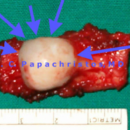
1. Blue arrows: the resected osteochondroma
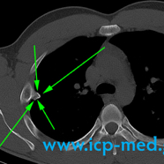
3. Preop CT: green arrows: tumour
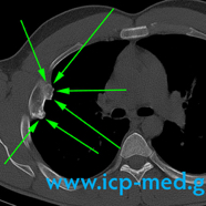
4. Preop CT: the irregularly–shaped tumour
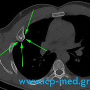
5. Preop CT
6. Preop CT
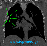
7. Preop MRI (coronal view)
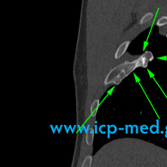
8. Preop MRI (sagittal view)
9. Preop MRI (sagittal view)
11. The resected tumour (green arrows)